Anterior Segment Ultrasound Machine
ArcScan Insight® 100 Device
The ArcScan Insight® 100 is an intelligent anterior segment imaging system that uses very high frequency ultrasound technology. The ArcScan Insight® 100 device and software create a powerful platform that enables users to easily obtain stunning high resolution images in micron precision of the entire anterior segment, including areas that can not be imaged with optical technologies.

The ArcScan Insight® 100 device is a precision high frequency ultrasound device for imaging and biometry of the eye.
Our Swept Beam Liquid Interface™ technology delivers dramatic developments in ultrasound imaging not seen in decades!
It uses core proprietary technology that originally developed in cooperation with Columbia/Weill University. A mechanically controlled 50 MHz transducer scans the eye in an arc shaped motion whereby its curvature approximates the anterior ocular surface.

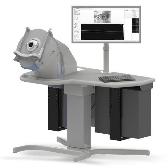
The device is designed to comfortably position the patient so their eye is looking through the center of the EyeSeal Disposable while keeping their head still. EyeSeal Disposables are used to prevent cross contamination between patients.
Designed for functional efficiency and ease of use, the system is comprised of four main components: the ArcScan Insight® 100 Device , the Insight software package, the fluidics module and the electronics unit.
CLINICAL BENEFITS INCLUDE
Precision imaging behind the iris for better ICL/IOL sizing
Comprehensive anterior segment measurement tracking for early glaucoma management and MIGS evaluation
Wide-angle cornea layer mapping for keratoconus screening and inlay evaluations

The ArcScan Insight® 100 is not a handheld device, yet it utilizes many of the components of modern optical imaging systems.
With patient seat times of just a few minutes, the device produces images with 1 micron resolution of the entire anterior segment: from the cornea, to the posterior of the lens.
In addition, measurements can also be made of the anatomic structures comprising the anterior of the eye such as anterior chamber depth, behind the iris, angle-to-angle width, and sulcus-to-sulcus width, and pathologic structures, such as solid masses and cysts.